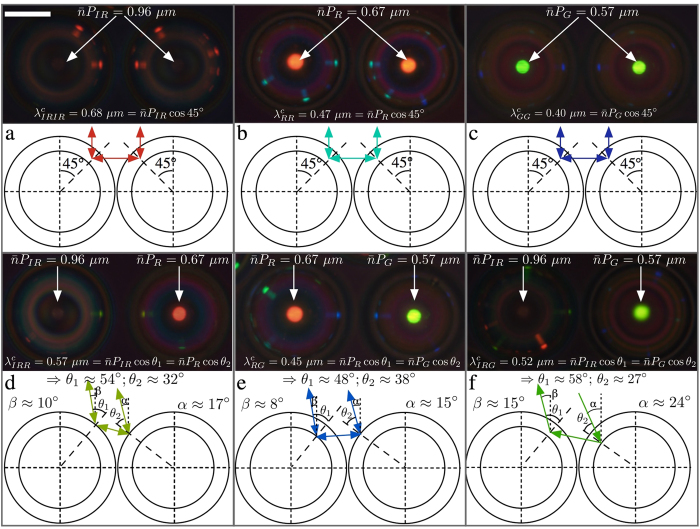
Figure 5.
Photonic cross communication between shells with identical (a–c) and different (d–f) helix pitches. The top part of each pane shows the reflection polarising microscopy texture together with the equations governing the reflection wavelengths (determined spectrophotometrically, see Supporting Information), while the schematics underneath illustrate communication pathways. The scale bar in (a) is 100 μm. No TIR spots appear since the sample is studied within a glass capillary.

