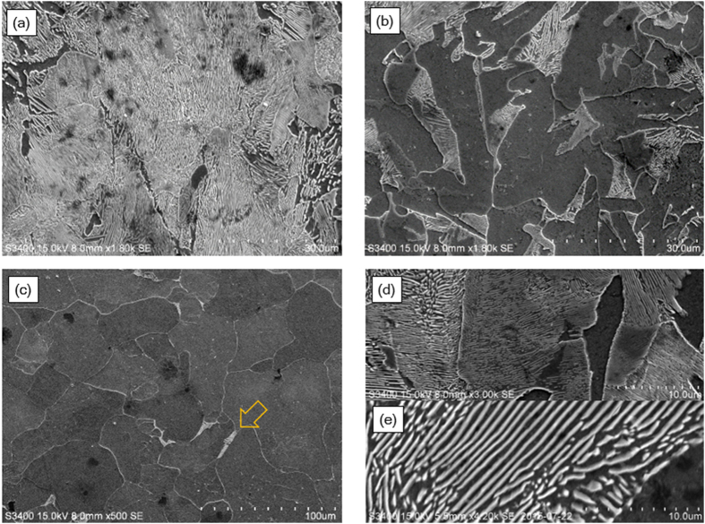
Figure 2.
SEM images of (a–c) C3H-AC and (d,e) lamella spacing variations of (d) C3H-AC and, (e) C3H-FC. For C3H-AC, the images are obtained at the different points: (a) 50 μm, (b) 300 μm from the surface, and (c) the center. Pearlite phase was also found in the center region as indicated by an arrow.

