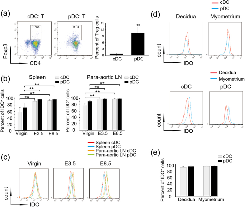
Figure 3. PDCs are more effective than cDCs at Treg induction independent of IDO expression.
(a) CDCs and pDCs were sorted from the para-aortic LNs of gravid mice, and CD4+ CD25− T cells were sorted from the spleens of OTII mice. Different DC subsets were co-cultured with CD4+ CD25− T cells for 7 days, and CD4+ CD25+ FoxP3+ Treg cells were detected by flow cytometry. Left panel-Representative FACS profiles are shown; Right panel- Histogram displays the statistical results of three independent experiments. (b) Virgin mice and allogeneic mated female C57BL/6 mice were sacrificed on E3.5 or E8.5, and IDO+ populations of cDCs and pDCs from the spleens and para-aortic LNs of four mice were analyzed. (c) Representative flow cytometry depicting MFI values for IDO expression in the spleens and para-aortic LNs on the indicated days from three independent experiments. (d,e) Cell suspensions were prepared from the separated decidua and myometrium of gravid mice at E8.5. The data summarize the percentages of IDO+ cDCs and pDCs (d) and the MFI value of IDO in the two subsets in the decidua and myometrium (e). The data represent the mean ± SD of three independent experiments from at least three mice. *P < 0.05; **P < 0.01.

