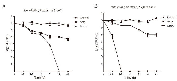
Figure 1.
Time-kill experiment of LBDv for Escherichia coli and Staphylococcus epidermidis. Y-axis represents the logarithm of colony forming units (CFU) determined by serial dilution on LB agar. X-axis represents the time point after incubation with 64 μM LBDv peptides. The 64 μM ampicillin and pGFP peptides are used as positive and negative control. Data are shown by the means ± S.E. Three replicate experiments are performed.

