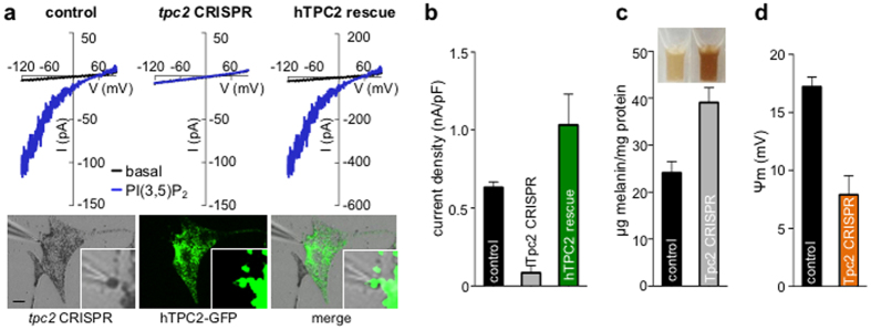
Figure 4. IPIP2 is mediated by TPC2.
(a) Representative I-V relationships of currents recorded before (black) or during (blue) application of 2 μM PI(3,5)P2, from melanosomes dissected from control Oa1−/− melanocytes, tpc2-targed CRISPR-Cas9 (tpc2 CRISPR), or tpc2 CRISPR rescued with human TPC2-GFP. Images: Representative rescue patch-clamp experiment from a melanosome expressing TPC2-GFP dissected from a cell expressing tpc2-targed CRISPR-Cas9 (scale bar = 10 μm). (b) Average current densities (nA/pF) measured at −120 mV (n = 3–9 melanosomes per condition, p < 0.0001 for tpc2 CRISPR vs. control or rescue). (c) Melanin content was increased in melan-a melanocytes expressing tpc2-targeted CRISPR-Cas9 compared with control melanocytes (n = 3 experiments, p < 0.01 for control vs. tpc2 CRISPR). Inset: Solubilized melanin from an equal number of control or tpc2 CRISPR cells. (d) In the presence of 2 μM PI(3,5)P2, average resting melanosomal membrane potential (Ψm) was significantly higher in melanosomes from control vs. tpc2 CRISPR Oa1−/− melanocytes (n = 3–4 melanosomes per condition, p < 0.01 for control vs. tpc2 CRISPR).

