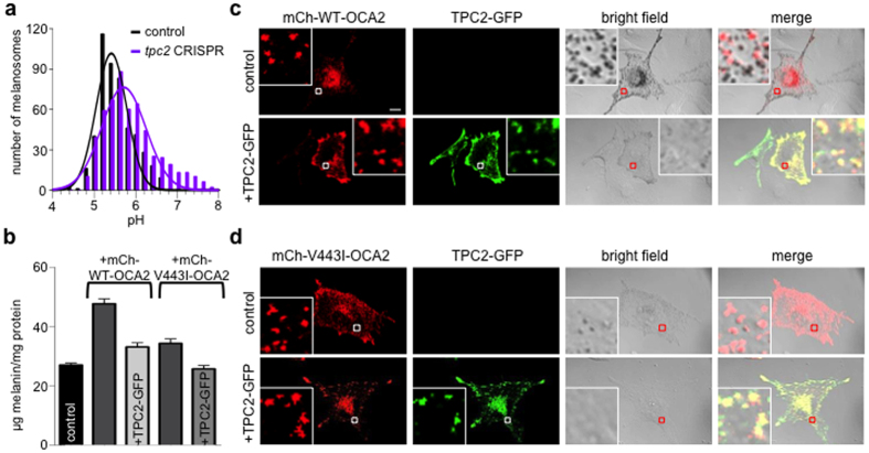
Figure 5. TPC2 regulates melanosomal pH to modulate pigmentation.
(a) Histogram of pH measurements of melanosomes from B16-F1 melanocytes expressing mCherry (mCh)-tagged V443I OCA2 and control or tpc2-targeted CRISPR-Cas9 fitted to Gaussian distributions. n = 496–543 melanosomes from 3 independent experiments, p < 0.0001 for average pH in control (5.51 ± 0.02) vs. tpc2 CRISPR (5.85 ± 0.03). (b) Melanin content (μg melanin/mg protein) of B16-F1 melanocytes (control, 27.0 ± 0.6) was significantly increased by expression of mCh-WT-OCA2 (47.6 ± 3.1, p < 0.001 compared to control) and decreased by coexpressing TPC2-GFP (33.1 ± 1.4, p < 0.01 compared to mCh-WT-OCA2 expressing cells). Expression of mutant mCh-V443I-OCA2 also increased melanin content compared to control B16-F1 melanocytes (34.3 ± 1.5, p ≤ 0.01) and this effect was reversed by coexpressing TPC2-GFP (25.7 ± 1.2, p < 0.01 compared to mCh-V443I-OCA2 expressing cells) (± s.e.m., n = 3 experiments). (c) In B16-F1 melanocytes mCherry-tagged wild-type (WT) OCA2 (red) induces pigmentation that is markedly reduced by coexpression with TPC2-GFP (green). Enlarged images of outlined regions shown in lower panels. Scale bar = 10 μm. (e) Expression of mCherry-tagged V443I mutant OCA2 (red) in B16-F1 cells induces reduced pigmentation that is further reduced by coexpression with TPC2-GFP (green).

