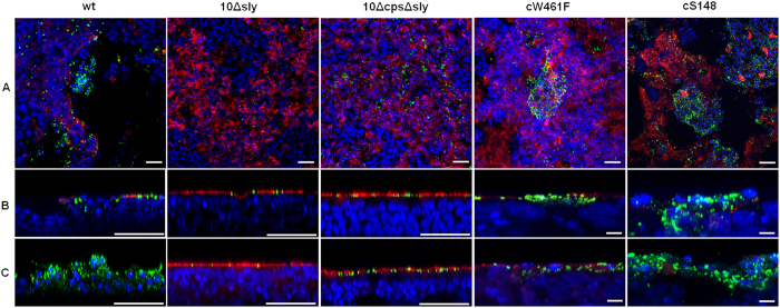
Figure 5. Suilysin-dependent damage of PBEC after infection with S. suis.
PBEC were apically infected with approximately 1 × 10⁷ CFU of S. suis wt, 10Δsly, 10ΔcpsΔsly, cW461F or cS148 respectively. After 4 h, cells were washed thoroughly to remove non-adherent bacteria, and further incubated under air-liquid interface conditions until 48 hpi (A,B) and 96 hpi (C). For analysis by confocal laser scanning microscopy, immunostaining was performed to visualize cilia (red) and streptococci (green). Nuclei were stained by DAPI (blue). Bars represent 25 μm in horizontal sections (A) and 50 μm (larger bar) or 5 μm (short bar) in vertical sections (B,C). Experiments were performed at least three times.

