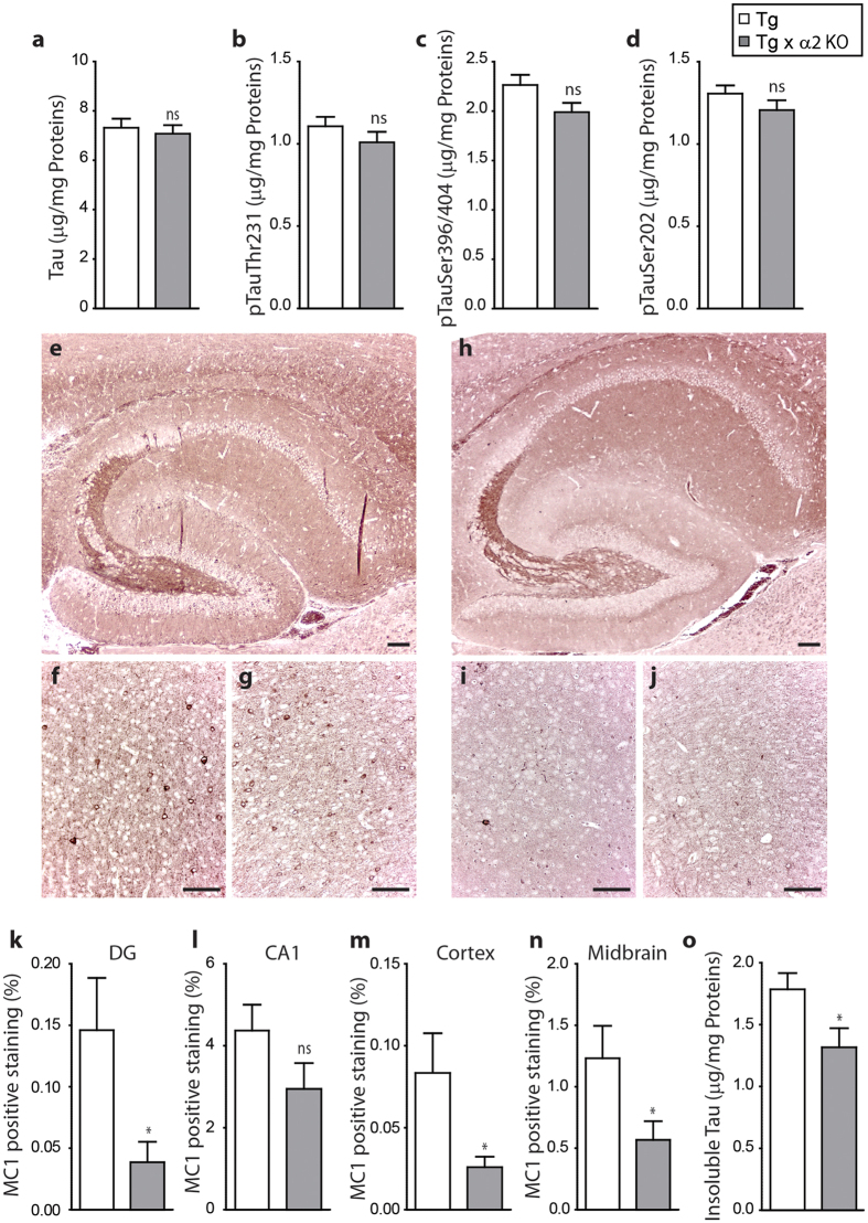
Figure 7. Impact of AMPK α2 deficiency on tau pathology.
(a–d) ELISA quantifications of total tau (a) and phosphorylated tau at epitopes Thr231 (b), Ser396/404 (c) and Ser202 (d) in heat stable fraction of 8-months old AMPKα2+/+: tauP301S (Tg) and AMPKα2−/−:tauP301S (Tg × α2 KO) mouse brains. (e–j) Immunohistochemistry of MC1 in Tg (e–g) and Tg × α2 KO (h–j) mouse brains in hippocampus (×5) (e,h), cortex (×20) (f,i) and midbrain (× 20) (g,j). Scale bars = 100 μm. (k–n) Quantifications of MC1 positive staining in dentate gyrus (DG, k) CA1 (l) cortex (m) and midbrain (n) expressed in percentage. Results represent mean ± SEM, n = 6, *p < 0.05 (Student’s t test). (o) ELISA quantifications of total tau in insoluble fraction of Tg and Tg × α2 KO mouse brains. Results represent mean ± SEM, n = 22, *p < 0.05 (Student’s t test).

