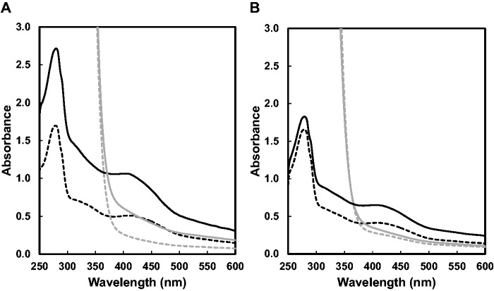
Fig. 2.
UV-Vis absorption spectra of the as-purified and reconstituted wild-type and C32S mutant of PqqE-N. (A) UV-Vis absorption spectra of the as-purified (black broken line) and reconstituted (black solid line) wild-type PqqE-N are shown with those 20-min after reduction with excess sodium dithionite (grey solid and broken lines). (B) UV-Vis absorption spectra of the as-purified (black broken line) and reconstituted (black solid line) C32S mutant of PqqE-N are shown with those 20-min after reduction with excess sodium dithionite (grey solid and broken lines). Enzyme concentrations were all adjusted to 1 mg/ml in buffer A.

