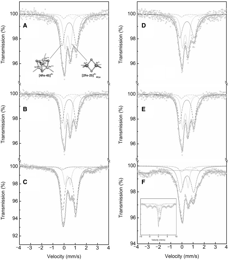
Fig. 3.
Mössbauer spectra of the wild-type and C32S mutant of PqqE-N. 57Fe Mössbauer spectra of (A) the as-purified wild-type PqqE-N, (B) the as-purified C32S mutant of PqqE-N, (C) the reconstituted wild-type PqqE-N, (D) the as-purified wild-type PqqE-N exposed to O2 for 1 day, (E) the as-purified C32S mutant of PqqE-N exposed to O2 for 1 day and (F) the reconstituted wild-type PqqE-N exposed to O2 for 1 day, all recorded at 5 K. In (A–F), a wide doublet (dashed line) is the simulated spectrum for [4Fe-4S]2+ cluster, a narrow doublet (dotted line) is the simulated spectrum for [2Fe-2S]2+4Cys cluster, small doublets (grey dashed and dotted lines) are those for the two minor unassigned doublets having hyperfine parameters identical with those reported for the [4Fe-4S]2+-pyruvate complex (48), a broad doublet (grey solid line) and the distributed sexted (grey solid line) are those for the free (non-cluster) high-spin Fe2+ and ferrihydrite (hydrous ferric oxyhydroxide), respectively, and black solid line is the sum of these simulated spectra. In (A), structure models for [4Fe-4S]2+ and [2Fe-2S]2+4Cys clusters drawn by PyMOL (Schrödinger, LLC) are shown. In (F), the spectrum covering the whole sexted of ferrihydrite is shown in a wider velocity range (inset).

