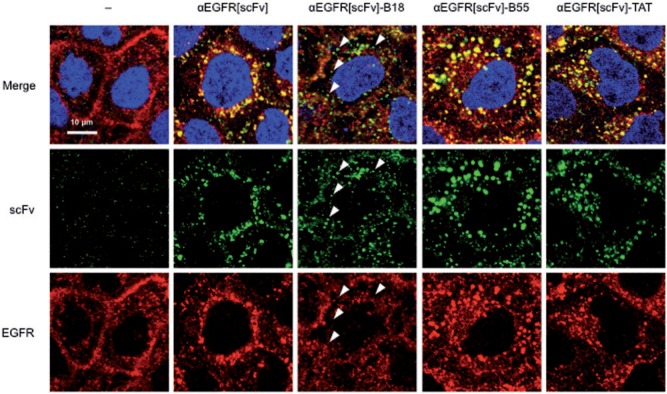
Fig. 2.
Confocal microscopic images of A431 cells treated with FP-fused αEGFR scFv proteins. The localization patterns of FP-fused αEGFR[scFv] proteins (300 nM each) and of EGFR in A431 cells were detected by immunocytochemistry using CF488-conjugated IgG (green) for scFv and Alexa 568-conjugated IgG (red) for EGFR. Cells were also stained with DAPI (blue). Arrowheads indicate small particles containing αEGFR[scFv]-B18 but not colocalized with EGFR.

