
FIG. 4.
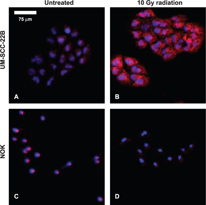
A mosaic of slides, stained for HIF-1α (red) and nuclei (blue) in vitro, of NOK and UM-SCC-22B, both treated with 10 Gy doses and untreated. These images do not represent the same field of view. A minimum size threshold for objects of interest removed the speckle from image quantification.