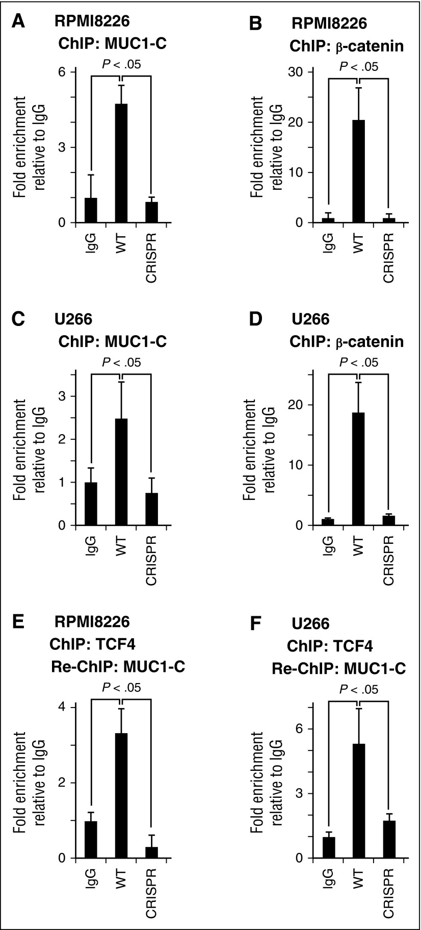
Figure 4.
MUC1-C occupies the MYC promoter. (A-D) Soluble chromatin from the indicated WT and CRISPR cells was precipitated with anti-MUC1-C (A,C), anti-β-catenin (B,D), or a control IgG. (E-F) In re-ChIP studies, anti-TCF4 precipitates were released and reimmunoprecipitated with IgG or anti-MUC1-C. The final DNA samples were amplified by qPCR with pairs of primers (supplemental Table 2) for the TBS in the MYC promoter. The results (mean ± SD of 3 determinations) are expressed as the relative fold enrichment compared with that obtained for the IgG control (assigned a value of 1).

