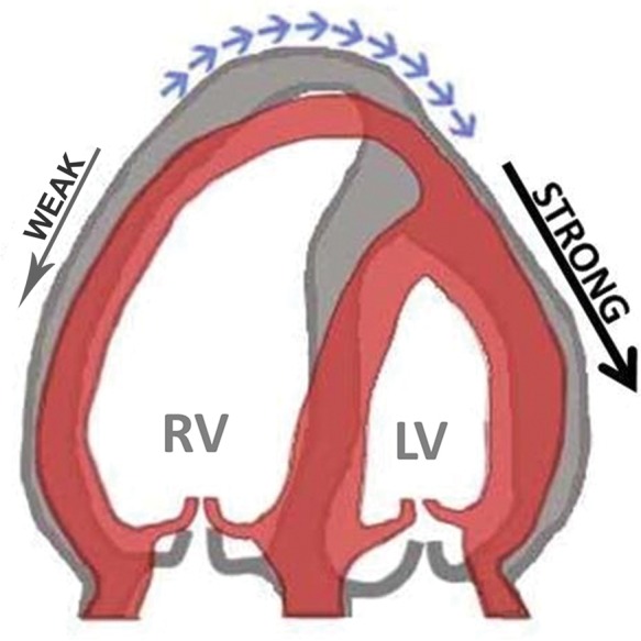
Figure 1.

Schematic of the mechanism of apical traction as it can be observed in the apical four-chamber view. Impaired contraction of the RV and traction from the normally contracting LV results in abnormal motion of the cardiac apex towards the left (blue arrows). Grey, diastole; red, systole.
