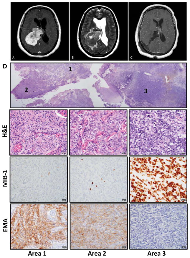
Figure 1.
Intratumoral heterogeneity within a high-grade meningioma. (A) T1-weighted gadolinium-enhanced axial MRI demonstrating a heterogeneously appearing contrast-avid tumor within the atrium of the right lateral ventricle, with (B) significant peritumoral edema and associated hydrocephalus on the T2-weighted axial MRI. (C) Post-operative T1-weighted axial MRI demonstrating complete resection. (D) Section of tumor, with 3 distinct appearing histologic regions (designated areas 1, 2, 3). Immunohistochemistry for epithelial membrane antigen (EMA) and MIB-1 (Ki-67). Scale bar, 10 μm.

