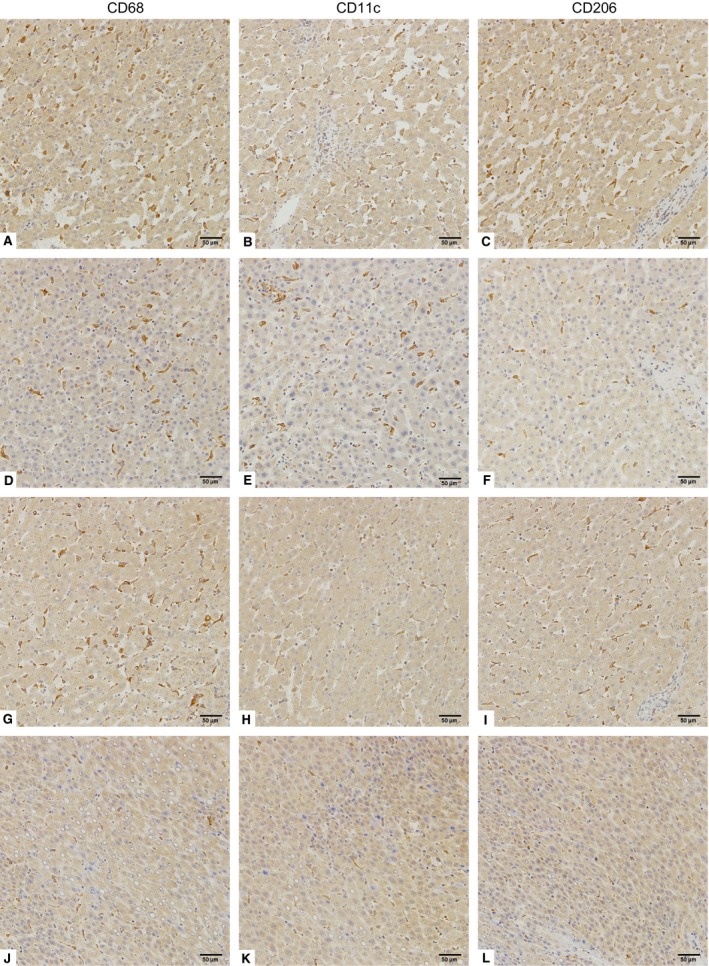
Figure 1.

Polarized macrophage infiltration in HCC tissues. Representative images of CD68 (A, D, G and J), CD11c (B, E, H and K) and CD206 (C, F, I and L) immunohistochemical staining in HCC tissue (original magnification ×200). (A–C) show high densities of CD68‐, CD11c‐ and CD206‐positive macrophages. (D–F) show high densities of CD68‐ and CD11c‐positive macrophages, but low CD206‐positive macrophage density. (G–I) show high densities of CD68‐ and CD206‐positive macrophages, but low CD11c‐positive macrophage density. (J–L) show low densities of CD68‐, CD11c‐ and CD206‐positive macrophages.
