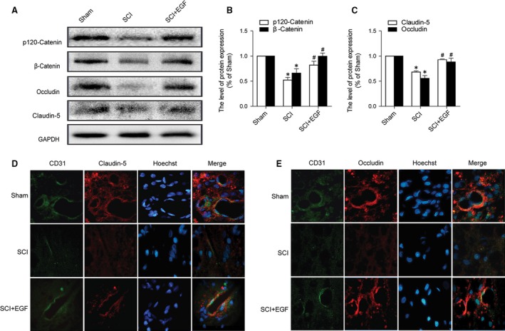
Figure 2.

EGF administration prevents the degradation of TJ and AJ proteins after SCI. (A) Protein expressions of p120‐Catenin, β‐Catenin, Occludin and Claudin‐5 in the spinal cord segment at the contusion epicentre. GAPDH was used as the loading control and for band density normalization. (B and C) The optical density analysis of p120‐Catenin, β‐Catenin, Occludin and Claudin‐5 protein, *represents P < 0.05 versus the Sham group, #represents P < 0.05 versus the SCI group, n = 5. (D and E) Double immunofluorescence shows that TJ and AJ proteins colocalize in CD31 (endothelial cell marker)‐positive blood vessels in the Sham, SCI rat and SCI rat treated with EGF groups.
