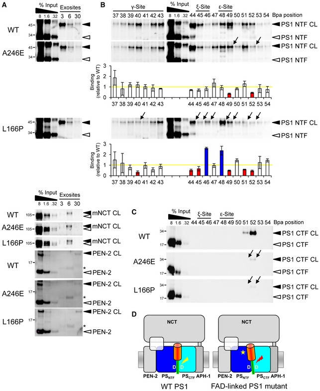
Figure 5. Mispositioning of the substrate cleavage domain by PS1 FAD mutations.

- PS1 FAD mutations display little or no effect on substrate interactions at the exosites. Asterisks indicate antibody crossreactivities to C99 monomer.
- PS1 FAD mutations alter the substrate binding to the PS1 NTF in the γ‐secretase cleavage domain. The weak PS1 A246E FAD mutation alters substrate interactions predominantly at V49 and L52, whereas the highly pathogenic PS1 L166P FAD mutation causes more broad changes over the whole cleavage domain. Arrows denote the changes in binding of individual residues. Corresponding quantitation of substrate binding is shown below the immunoblots. Bars denote the mean ± SE (n = 3). Yellow line indicates the level of substrate binding to PS1 WT, which was set to 1. Blue and red bars indicate the increases or decreases in binding, respectively.
- Both mutants decrease crosslinking to the PS1 CTF.
- Model depicting substrate mispositioning of the PS1 FAD mutants in the active site region. Red lightning mark indicates corresponding substrate miscleavage.
