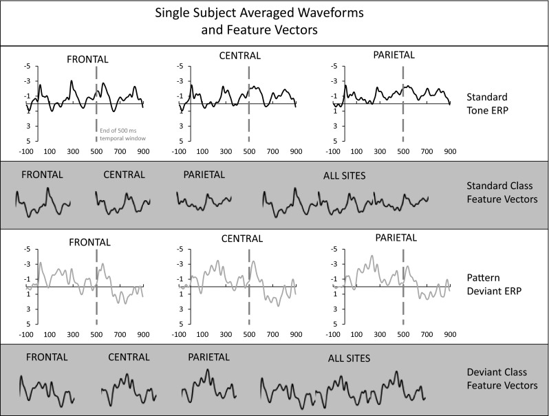
Fig. 2.

An illustration of averaged waveforms and feature vectors for a single subject. The upper panel shows standard tone averaged waveforms at the three scalp sites (Frontal, Central, and Parietal) using all available epochs. All amplitudes are in microvolts and all time values are in milliseconds (horizontal axes). The end of a 500-ms temporal window is delimited by a gray dashed line. The middle upper panel demonstrates how the data from that temporal window are used to create Frontal, Central, Parietal, and all scalp sites feature vectors for the standard ERP class. The lower middle panel shows pattern deviant averaged waveforms. Again, a 500-ms temporal window is delimited, and the composition of the Frontal, Central, Parietal, and all scalp sites feature vectors are shown, now for the deviant ERP class, in the bottom panel
