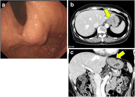
Fig. 1.

a Gastroduodenal endoscopy revealing the presence of a submucosal tumor at the greater curvature of the esophagogastric junction. b, c The enhanced computed tomography (CT) scan shows a smooth-outlined hypervascular solid mass (23 mm × 20 mm) in the gastric wall at the esophagogastric junction (arrow)
