
Extended Data Fig. 6. Majority of CD4 T cells recruited to the DRG and spinal cord of immunized mice is localized in the parenchyma of neuronal tissues.
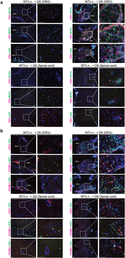
(a) C57/BL6 mice were immunized i.n. with TK− HSV-2. Six days after challenge of immunized mice six weeks prior, neuronal tissue sections (DRG and spinal cord) were stained for CD4+ cells and VCAM-1+ cells or CD31+ cells (red or green). Blue labeling depicts nuclear staining with DAPI (blue). Images were captured using a 10× or 40× objective lens. Scale bars indicate 100 µm. (b) C57/BL6 mice were immunized i.n. with TK− HSV-2. Six weeks later, mice were challenged with WT HSV-2 ivag and neuronal tissues were collected 6 days later. DRG and spinal cord were stained for CD4+ cells (red) and MHC class II+ cells, CD11b+ cells or Ly6G+ cells (green). Blue labeling depicts nuclear staining with DAPI (blue). Images were captured using a 10× or 40× objective lens. Scale bars indicate 100 µm. These data are representative of at least three similar experiments.