
Extended Data Fig. 9. Vascular permeability in DRG and spinal cord is augmented following WT HSV-2 challenge.
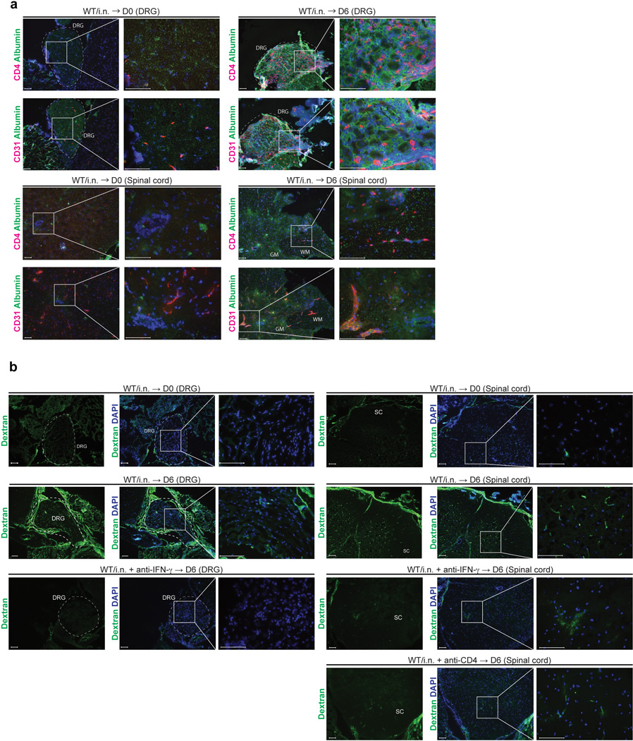
(a) C57/BL6 mice were immunized i.n. with TK− HSV-2. Six days after challenge of immunized mice six weeks prior, neuronal tissue sections (DRG and spinal cord) were stained for CD4+ cells (red) and mouse albumin (green). Blue labeling depicts nuclear staining with DAPI (blue). (b) C57/BL6 mice were immunized i.n. with TK− HSV-2. Six weeks later, these mice were challenged with lethal WT HSV-2. Six days after challenge, Oregon green 488-conjugated dextran (70 kDa) (5 mg/ml, 200 µl/mouse) was injected i.v. into intranasal immunized mice. Forty five minutes later, these mice were sacrificed for immunohistochemical analysis. GM; gray matter, WM; white matter. These data are representative of three similar experiments.