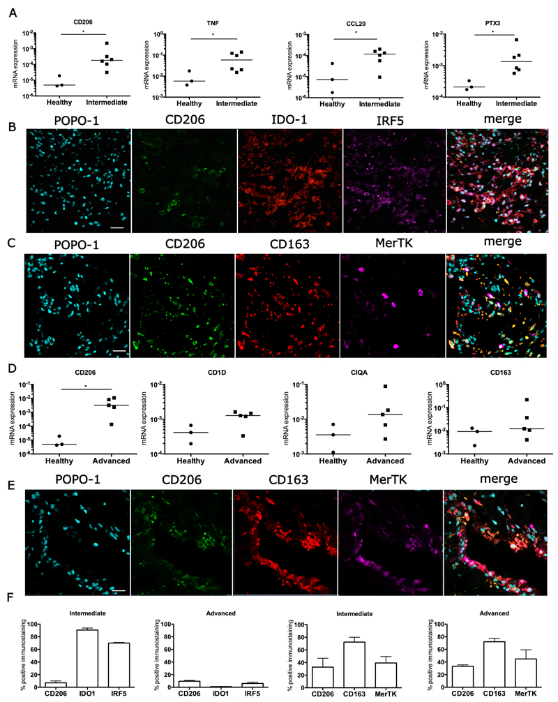
Figure 3.
Inflammation activation pathway signatures in intermediate and advanced stage disease tendons. (A) A mixed inflammation activation gene signature in intermediate stage diseased supraspinatus tendon samples (n=6) compared to healthy subscapularis tendons (n=3). (B, C) Representative immunofluorescence images of the inflammation activation protein signature in intermediate stage diseased tendons. (B) Markers indicate activation of the following pathways: STAT-6 (CD206, green), NF-κB (IDO-1, red), and IFN (IRF5, purple). (C) Markers indicate activation of the glucocorticoid receptor pathway (CD163, red); also shown is the tissue resident macrophage marker Mer tyrosine kinase (MerTK, purple). (D) A STAT-6/glucocorticoid receptor gene signature predominates in advanced stage diseased supraspinatus tendons (n=5) compared to healthy subscapularis tendons (n=3). Gene expression is normalized to β-actin. Statistically significant differences were calculated using pairwise Mann Whitney U tests. Bar represents median, *denotes p<0.05. (E) Representative immunofluorescence images of the inflammation activation protein signature in advanced stage diseased supraspinatus tendon. Shown is staining for the STAT-6 (CD206, green) and glucocorticoid receptor (CD163, red) activation pathways; tissue resident macrophages are stained with Mer tyrosine kinase (MerTK, purple). Cyan represents POPO-1 nuclear counterstain. Scale bar = 20µm. (F) Quantitative analysis showing the percentage of cells staining immunopositive for inflammation pathway proteins. Data are shown as mean and SEM.

