
Figure 1.
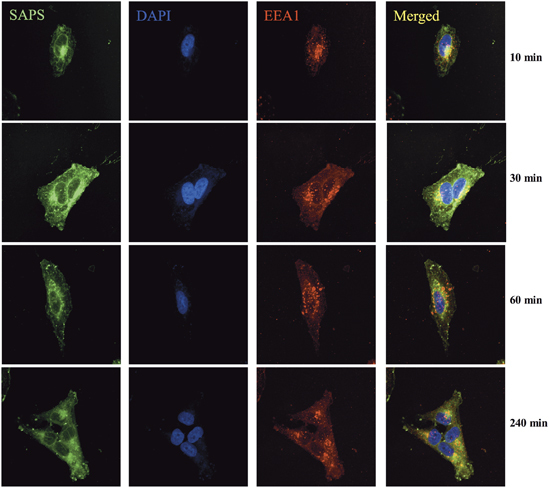
Endosomal localized 1-stearoyl-2-arachidonoyl-sn-glycero-3-phospho-L-serine (SAPS). BEAS-2B cells were incubated with TopFluor-SAPS (100 μg ml−1) for indicated time periods. Intracellular distribution of SAPS (green) partially colocalized within endosomes (EEA1/red). Images were collected using a Nikon confocal microscope and are representative of three independent experiments. The merged image shows overlay of areas positive for SAPS and endosome.