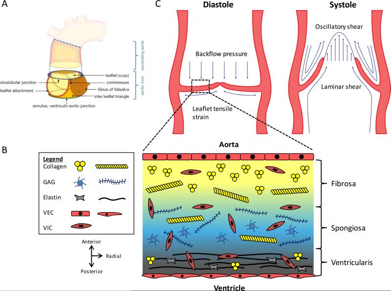
Figure 1.
Functional anatomy and heterogeneous composition of the aortic root. (A) The aortic root is a complex structure consisting of the leaflets and other structures, including the Sinus of Valsalva, commissure, sinutubular junction, ventriculo-aortic junction, and leaflet attachment. Figure from 12 and reprinted with permission. (B) Movat depiction of the ECM componets within the three layers of the aortic leaflet. Collagen is found throughout the valve, but it is packed tightly and aligned circumferentially in the fibrosa. Elastin is radially-aligned and predominately exists in the ventricularis. GAGs are mainly found in the spongiosa. Endothelial cells line the fibrosa and ventricularis sides, and valvular interstitial cells are found throughout the valve. (C) Biomechanical forces acting on the aortic root. During diastole, leaflets are stretched to form the coaptation and prevent backflow. Leaflets experience tensile strain and stress from the aortic pressure. In systole, leaflets are flexed open and experience both oscillatory and laminar shear stresses. Figure adapted from 16.

