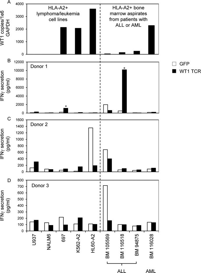
Figure 4.
WT1 RNA expression in tumor cell lines and bone marrow aspirates from HLA-A*0201+ patients with leukemia and recognition by PBL retrovirally transduced to express a WT1 reactive TCR. (A) Quantitative RT-PCRs for WT1 and GAPDH were conducted on RNAs isolated from tumor cell lines and bone marrow aspirates. WT1 RNA copies were divided by GAPDH copies and expressed as WT1 copy number per 1e6 GAPDH copy number. (B-D) PBL from 3 HLA-A*0201+ donors were transduced with retroviral particles encoding GFP as a negative control or with the WT1 reactive TCR (TRAV12-1*01; CDR3: CVVNTPPNTDKLIF; TRBV7-2*01; CDR3: CASTPFTSGSGWDEQFF). Transduced T cells were cocultured overnight with tumor cell lines and bone marrow aspirates. IFNγ in coculture supernatants was measured by ELISA. * indicates >100 pg/ml IFNγ secretion by WT1 TCR transduced T cells, >2X background with any negative control target cell, and >2X background of GFP transduced T cells with the same target cell.

