Figure 2.

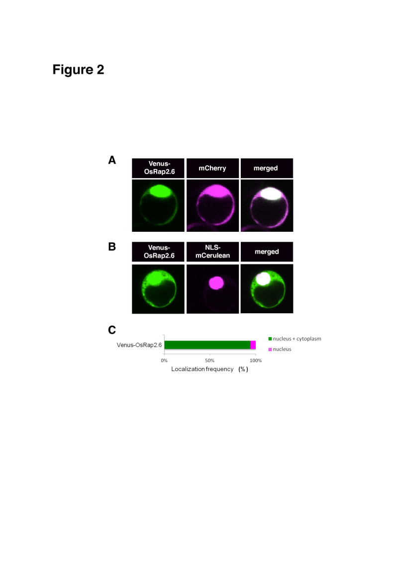
Subcellular localization of OsRap2.6. Rice protoplasts were transformed with known fluorescent proteins mCherry and NLS-mCerulean. Fluorescence was detected using a CCD camera connected to a confocal microscope. The localization frequency of the cells was analyzed in 50–100 cells expressing YFP/CFP as compared to the positive controls using Excel. Means and standard deviations were separated using Student’s t -test (p<0.05). (A) Subcellular localization of Venus-OsRap2.6 with the mCherry. (B) Subcellular localization of Venus-OsRap2.6 with the NLS-mCerulean. (C) Localization frequency of Venus-OsRap2.6.
