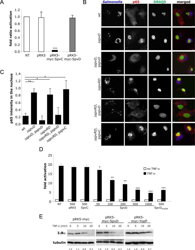
Fig 2. SpvD prevents nuclear accumulation of p65 but not degradation of IĸBα.
(A) SpvD does not inhibit activation of an AP-1-regulated promoter. HEK-293 cells were cotransfected with an AP-1-dependent luciferase reporter plasmid, pTK-Renilla luciferase and pRK5myc, pRK5myc-SpvC or pRK5myc-SpvD. Cells were then stimulated with PMA for 6 h. Firefly luciferase activity was normalized against Renilla luciferase activity. Results are expressed as fold induction compared to non-transfected (NT) cells. Values are expressed as mean ± SEM of 3 independent experiments and P-values were obtained using two-tailed unpaired Student's t-test (*** p < 0.005). (B-C) SpvD prevents nuclear translocation of p65. (B) Representative immunofluorescence fields of p65 localisation using anti-p65 (red) in TLR4 -/- BMMs infected for 10 h with indicated Salmonella strains (blue). Cell nuclei were stained with DRAQ5 (green). Scale bar, 8 μm. (C) Quantification above background levels of p65 intensity in the nucleus was analyzed by three-dimensional (3D) confocal microscopy. Background levels were determined using signals measured in uninfected cells. P-values were obtained using two-tailed unpaired Student's t-test (*p < 0.05; ** p < 0.01). (D) SpvD inhibits activation of an NF-ĸB -regulated promoter. HEK-293 cells were co-transfected with an NF-ĸB -dependent luciferase reporter plasmid, pTK-Renilla luciferase and pRK5myc, pRK5myc-SpvC, pRK5myc-SpvD or pRK5myc-SpvDK185A at the indicated amounts (in ng). The NF-ĸB pathway was activated with TNF-α and luciferase activity was measured after 8 h. Results are expressed as fold activation in relation to unstimulated and non-transfected (NT) cells. Values are expressed as mean ± SEM of at least 3 independent experiments and statistical significances were calculated using ANOVA followed by Bonferonni's multiple comparison test against NT cells (*p < 0.05; ** p < 0.01; *** p < 0.005). (E) Degradation of IĸBα is not affected by SpvD. HEK-293 cells transfected by pRK5myc-SpvD or pRK5myc-YopP were prepared at the indicated times after TNF-α stimulation and analysed by SDS-PAGE and immunoblotting with anti- IĸBα and anti-tubulin antibodies. Ratio of IĸBα normalised to unstimulated cells is indicated below immunoblots.

