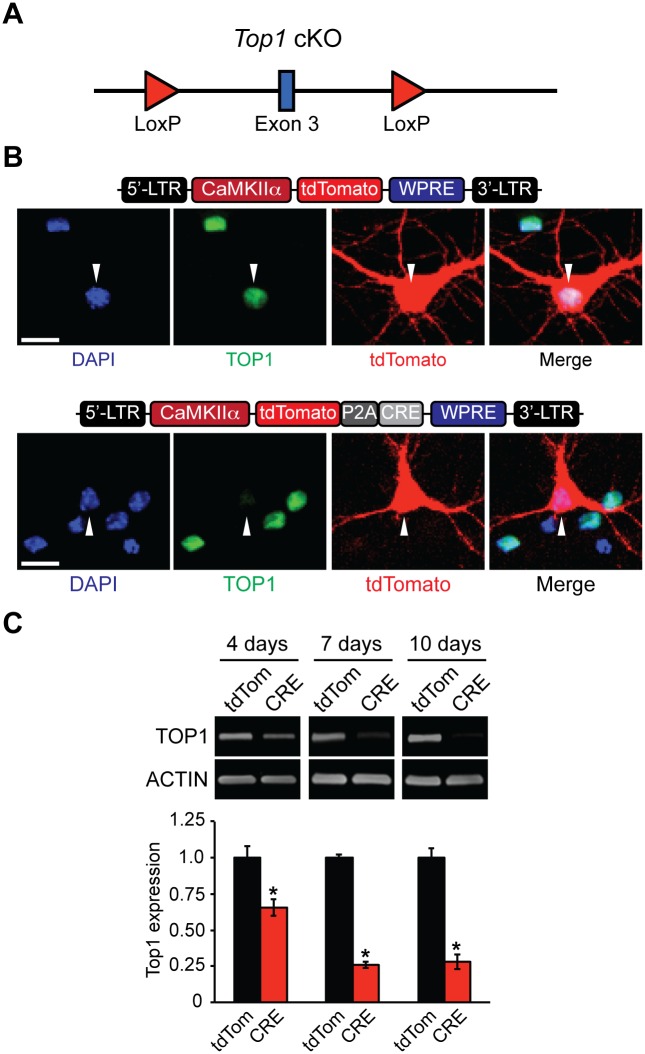
Fig 1. Generation and validation of Top1 cKO mouse.
(A) Schematic of the Top1 cKO allele. LoxP sites flank exon 3. (B) Schematic of tdTomato (top) and tdTomato-P2A-CRE lentiviral plasmids (bottom). Top1fl/fl neurons were transfected with tdTomato or tdTomato-P2A-CRE plasmids. Neurons were fixed and immunostained with an anti-TOP1 antibody. Scale bar, 10 μm. (C) Cortical neurons were infected with tdTomato or tdTomato-P2A-CRE lentivirus at DIV 3 and then were harvested at DIV 7, DIV 10, and DIV 13. Representative immunoblots and quantification of TOP1 protein expression normalized to ACTIN (bottom). Mean ± s.e.m., unpaired student’s t-test; * p < 0.05, n = 3 cultures.

