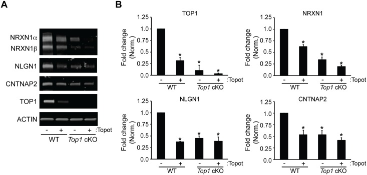
Fig 3. TOP1 deletion reduces synaptic adhesion protein levels.
(A) Top1fl/fl neuron cultures were infected with tdTomato (WT) or tdTomato-P2A-CRE (Top1 cKO) lentivirus at DIV 3. Cells were then treated at DIV 15 with vehicle (DMSO) or 300 nM topotecan for 72 hours. Shown are representative immunoblots with antibodies to NRXN1, NLGN1, CNTNAP2, TOP1, and ACTIN. (B) Quantification of fold change in TOP1, NRXN1, NLGN1, and CNTNAP2 protein expression normalized to ACTIN. Mean ± s.e.m., unpaired student’s t-test; * p < 0.05, n = 3 cultures.

