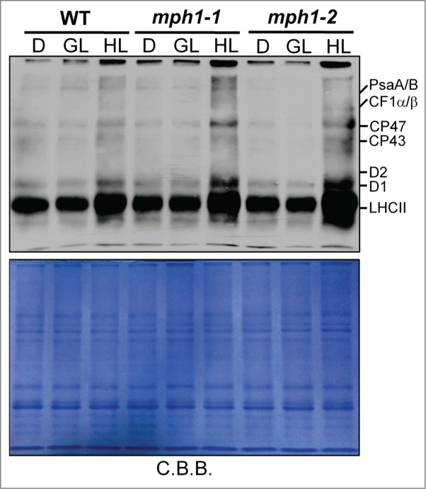
Figure 3.

Analysis of oxidation of thylakoid membrane proteins in mph1 mutants and wild-type plants. Leaves were harvested from plants after 16 h dark period (D), at growth light (GL) or after 3 h high-light treatment (HL). Thylakoids containing 2μg chlorophyll were loaded into each lane and carbonylated proteins in thylakoids were immunodetected using OxyBlotTM (Millipore). Shown below is the reference gel stained with Coomassie brilliant blue (C.B.B.).
