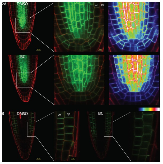
Figure 2.
I3C treatment affects auxin transporters. (A) Seedlings expressing PIN1:GFP were grown on MS medium for 5 days, treated with 200 μM of I3C or with DMSO for 30 minutes, and imaged using confocal microscopy (Zeiss LSM780, with a 40x water objective). Heat-maps represent GFP density. GFP fluorescence was quantified using ImageJ software. (B) Seedlings expressing PIN2:GFP were grown on MS medium for 6 days, treated with 200 μM of I3C or with DMSO for 30 minutes, and imaged using confocal microscopy (Zeiss LSM780, with a 40x water objective). Cell walls were stained using 0.005mg/ml propidium iodide. co = cortex, ep = epidermis.

