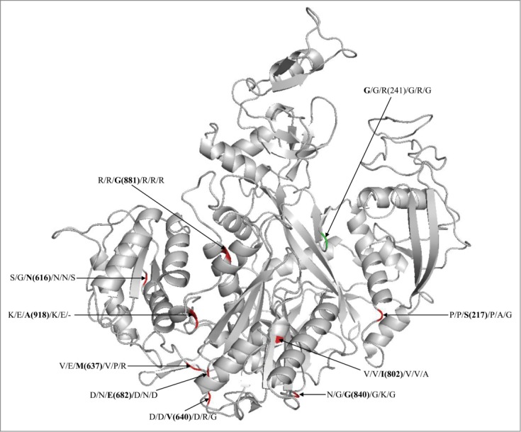
Figure 1.

Modeled structure of AtAGO4 showing the functionally diverged sites. The labeled residues separated with ‘/’ at each site are the residues from AtAGO1, AtAGO2, AtAGO4, AtAGO5, PptAGOLike1 and CrnAGO2, respectively. The value in parenthesis is the coordinate of residue in AtAGO4. The sites with red color indicates that it has diveged in AtAGO4 than the other AGOs, while with green color indicate divergence in AtAGO1 than the other AGOs.
