
Figure 7.
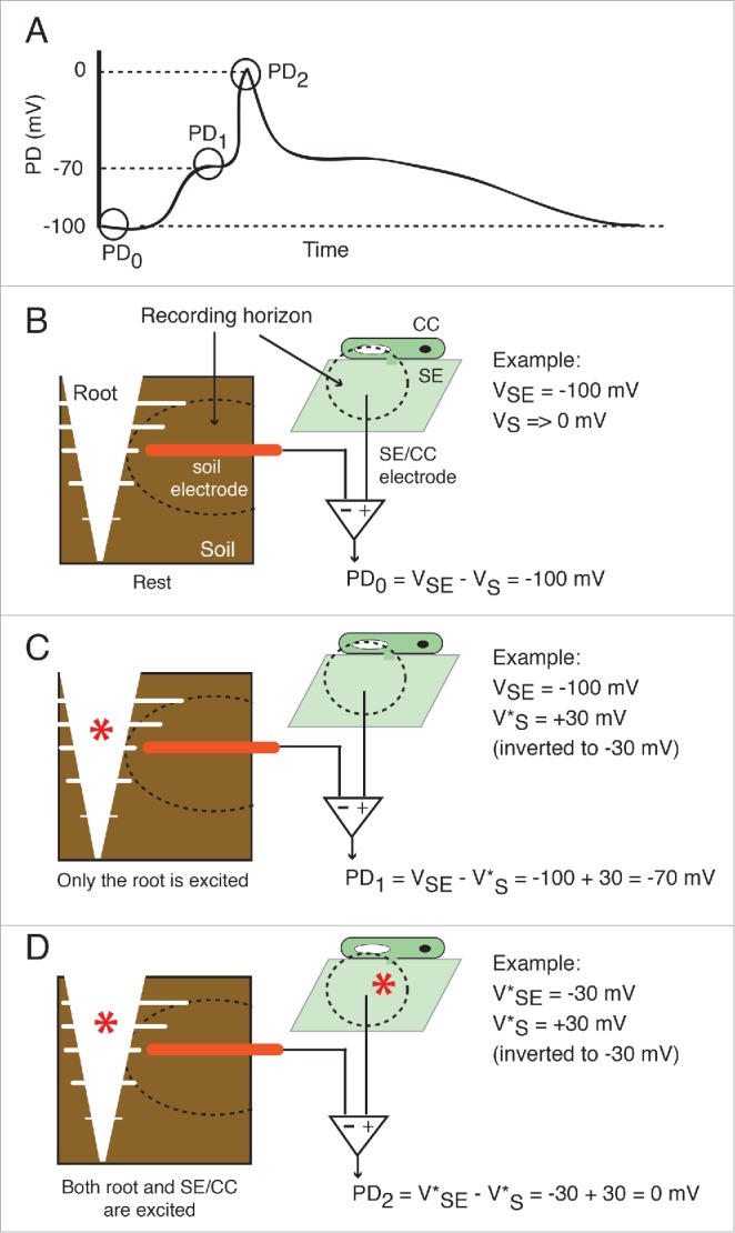
Proposed contributions of the root and the SE/CC network to the wound-induced action potential and long potential. (A) Drawn waveform of a typical wound-induced EPG-recorded systemic potential. The potential difference (PD) between the root and the SE/CC electrode is plotted as a function of time. In a typical EPG-recorded systemic signal there are 3 potential levels. (B) At rest (PD0) only the resting membrane potential of the SE/CC contributes to the PD. By convention, the extracellular or external resting potential (in this case external to the root) is regarded as negligible (Vs→0), although there are always background ionic currents. In the given example, the intracellular potential of the SE/CC has an arbitrary value of −100 mV. (C) The first potential level of a typical EPG-recorded signal (PD1) is generated by electrical activity in the root. At this point, the SE/CC is still unexcited. Intracellular depolarizations in root cells are recorded as a compound extracellular hyperpolarization by the root electrode. The size of the recorded extracellular hyperpolarization will be reduced proportionally with the distance between the root and the electrode. In the given example, the excitation (depolarization) of a root area is recorded externally as a +30 mV hyperpolarization (these are arbitrary values). Since the EPG uses a differential amplifier with an inverting input and a non-inverting input, the polarity of the root potential is reversed before calculating the PD. (D) The second potential level of a typical EPG-recorded signal (PD2) is generated by the still ongoing electrical activity in the root and a maximally excited SE/CC. In the given example, the maximal depolarization in the SE/CC is −50 mV. Abbreviations used are: SE = sieve element; CC = companion cell, VSE = membrane potential of the sieve element; VS = extracellular potential of the root, in the vicinity of the soil electrode. The excited depolarized membrane potential is shown with an asterisk (e.g. V*SE).