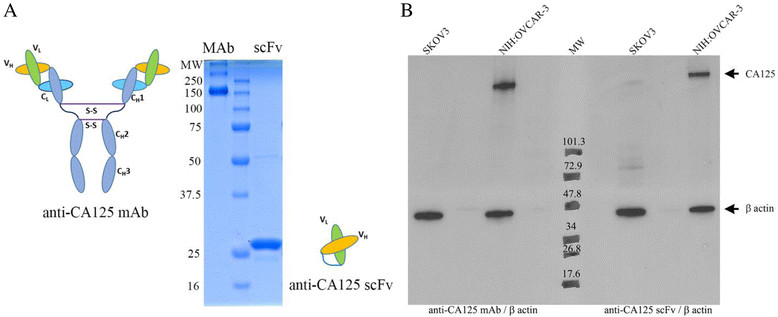
Figure 1.

Representative images and Western blots. (A) Representative images of purified anti-CA125 MAb (148 KDa) and scFv (28 KDa) upon SDS-PAGE under non-reducing conditions. (B) Western blots of SKOV3 (CA125-) and NIH:OVCAR-3 (CA125+) cells with anti-CA125 MAb, anti-β actin antibody (left side of molecular weight marker); anti-CA125 scFv, and anti-β actin antibody (right side of molecular weight marker).
