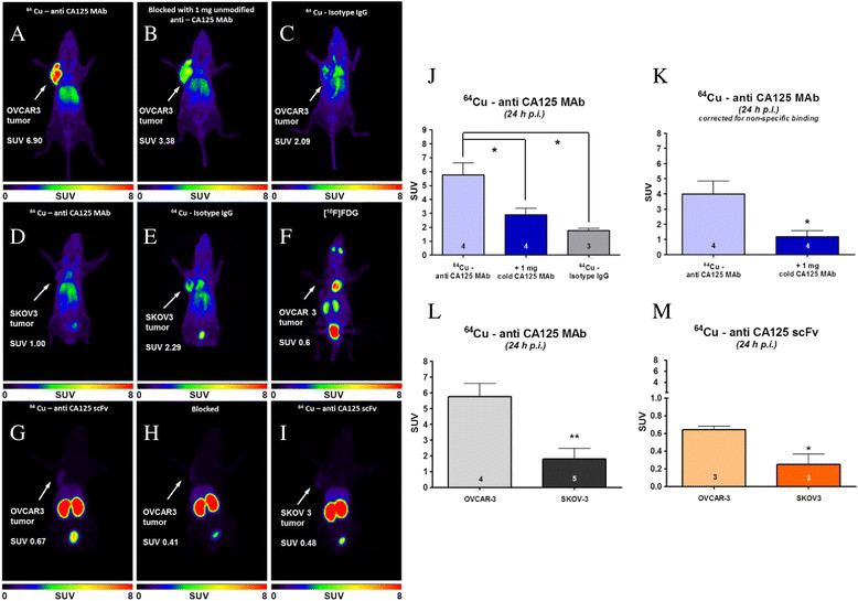
Figure 6.

PET images and diagrams. (A-E, G-I) Representative 24-h post-injection small-animal-PET images of xenograft mice injected with radioimmunoconjugates. (F) 1-h p.i. PET image of NIH:OVCAR-3 xenograft mouse injected with 18 F-FDG. Color intensity scale bars represent standardized uptake value (SUV) of radiotracer in animals. Diagrams on the right show (J) SUV in tumors of experimental and control animals using 64Cu-labeled anti-CA125 MAb, (K) SUV in NIH:OVCAR-3 tumors after correction for non-specific uptake, (L) tumor SUV obtained from injection of 64Cu-labeled anti-CA125 MAb in NIH:OVCAR-3 versus SKOV3 xenograft mice, and (M) tumor SUV obtained from injection of 64Cu-labeled anti-CA125 scFv in NIH:OVCAR-3 versus SKOV3 xenograft mice. Numbers of animals (n) are indicated at the bottom of each bar.
