
Figure 4.
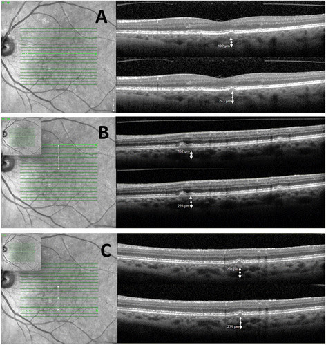
Patient with birdshot retinochoroidopathy. Spectral domain OCT of the left eye of a patient with birdshot retinochoroidopathy. The infrared SLO images (A to C) show the location of the horizontal b-scan. The correspondent b-scans show the vessel diameter and the choroidal thickness below the different lesions.