Figure 2.
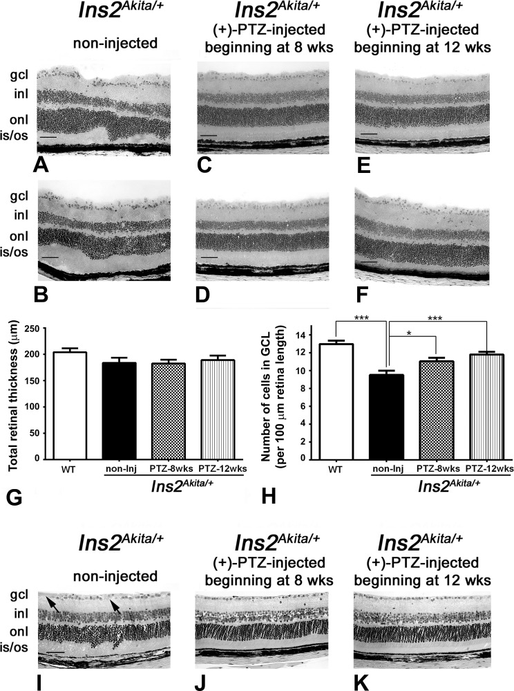
Preservation of retinal structure and cells in the GCL in Ins2Akita/+ mice administered (+)-PTZ after diabetes onset. All images are from mice age 25 weeks. (A, B) Representative H&E-stained retinal cryosections of Ins2Akita/+ mice that received no (+)-PTZ for the duration of the study; the INL is slightly disrupted, and GCL density is decreased. Representative H&E-stained retinal cryosections of (C, D) Ins2Akita/+ mice that received 0.5 mg kg−1 (+)-PTZ beginning 4 weeks after onset of diabetes (at age 8 weeks) or (E, F) Ins2Akita/+ mice that received 0.5 mg kg−1 (+)-PTZ beginning 8 weeks after onset of diabetes (at age 12 weeks). Retinas of (+)-PTZ injected mice demonstrated marked preservation of retinal structure. Morphometric analysis revealed no significant change in total retinal thickness in any of the eyes evaluated (G), but a significant difference in the number of cell bodies in the GCL per 100 μm length of retina (H). Eyes were processed for JB-4 embedding and stained with H&E to permit visualization of retinal architecture of (I) Ins2Akita/+ mice that received no (+)-PTZ; (J) Ins2Akita/+ mice that received 0.5 mg kg−1 (+)-PTZ beginning 4 weeks after onset of diabetes (at age 8 weeks); or (K) Ins2Akita/+ mice that received 0.5 mg kg−1 (+)-PTZ beginning 8 weeks after onset of diabetes (at age 12 weeks). Arrows in I point to areas of cell dropout. gcl, ganglion cell layer; inl, inner nuclear layer; onl, outer nuclear layer; is/os, inner/outer segments. Magnification bar: 50 μm. Data are means ± SE. Significantly different at *P < 0.05 and ***P < 0.001.

