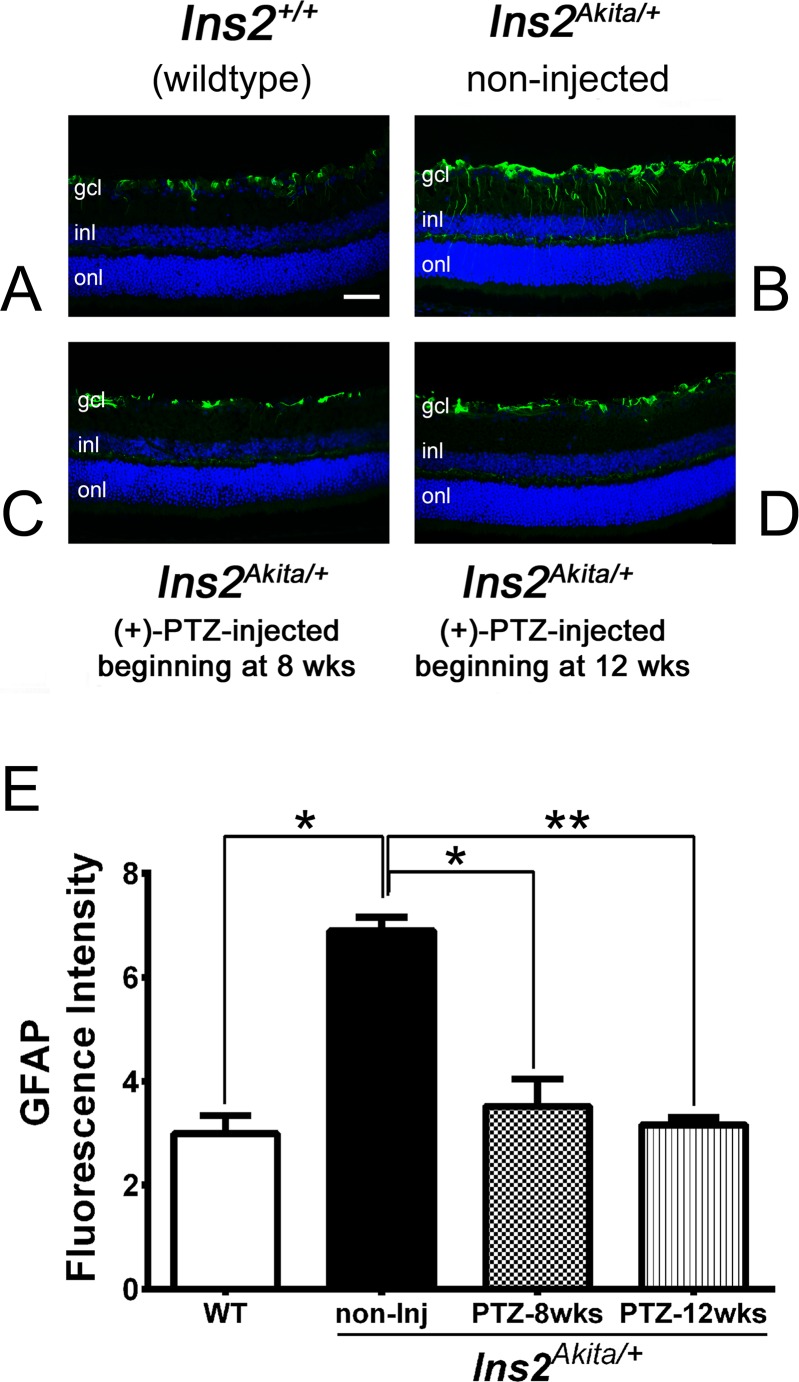
Figure 3.
Immunofluorescent detection of GFAP, a marker of gliosis in Ins2Akita/+ mice administered (+)-PTZ after diabetes onset. Representative photomicrographs of immunofluorescent detection of GFAP in retinal cryosections from (A) Ins2+/+ (WT), (B) Ins2Akita/+ [no (+)-PTZ treatment], (C) Ins2Akita/+ mice that received 0.5 mg kg−1 (+)-PTZ beginning 4 weeks after onset of diabetes (at age 8 weeks), and (D) Ins2Akita/+ mice that received 0.5 mg kg−1 (+)-PTZ beginning 8 weeks after onset of diabetes (at age 12 weeks). Immunofluorescence analysis used anti-GFAP followed by incubation with Alexa Fluor 488 (green-labeled secondary antibody), showing marked increase in GFAP in radially oriented fibers of Müller glial cells (radial labeling) in Ins2Akita/+ [no (+)-PTZ treatment], but minimal labeling in Müller cells of the WT or (+)-PTZ–treated mice. Astrocytic labeling with GFAP is typical and is observed in retinas of all mice. (E) Quantification of GFAP fluorescence intensity data obtained from metamorphic analysis (significant difference: *P < 0.05, **P < 0.01).

