Figure 4.
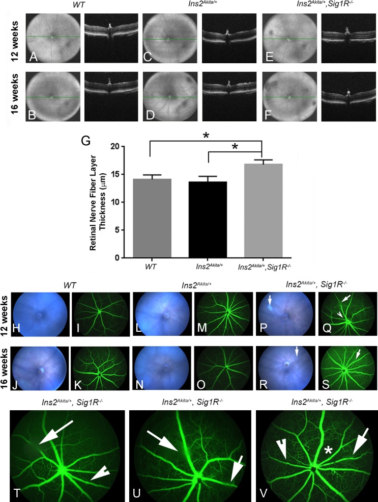
Spectral-domain OCT imaging, fundoscopy, and FA in Ins2Akita/+ and Ins2Akita/+/Sig1R−/−mice. Wild-type (Ins2+/+), Ins2Akita/+, and Ins2Akita/+/Sig1R−/− mice were anesthetized and subjected to SD-OCT to obtain images of the retina in situ. Representative VIP for WT (Ins2+/+) mice: age 12 (A) and 16 weeks (B); Ins2Akita/+ mice: age 12 (C) and 16 weeks (D); and Ins2Akita/+/Sig1R−/−mice: age 12 (E) and 16 weeks (F). The panel to the right of each VIP is the B-scans of the same animal. Autosegmentation analysis was performed to determine whether there were significant differences in thickness of retinal layers among the three mouse groups. There was a significant increase in thickness of the nerve fiber layer (G) (*significantly different at P < 0.05). Mice were subjected to fundoscopy WT (Ins2+/+) at 12 (H) and 16 weeks (J); Ins2Akita/+ at 12 (L) and 16 weeks (N); Ins2Akita/+/Sig1R−/− at 12 (P) and 16 weeks (R). Fundus images were normal for WT and Ins2Akita/+, whereas vitreal opacities (denoted by long arrows) were detected in the Ins2Akita/+/Sig1R−/− mice (P, R). Mice were subjected to FA to observe vascular filling with a fluorescent dye. Wild-type (Ins2+/+) showed no alterations at either 12 (I) or 16 weeks (K). Ins2Akita/+ mice also had normal vascularization patterns at 12 (M) and 16 weeks (O), whereas Ins2Akita/+/Sig1R−/− mice had beading at 12 (Q) and at 16 weeks (S). The long arrows in Q and S point to areas of vitreal opacity; the indented arrowhead points to a vessel with beading (S). (T) Enlarged image of image shown in S. (U, V) FA images of two additional mice at 16 weeks showing vitreal opacity (long arrow) and occasional beading of vessels (indented arrowhead). The asterisk in V denotes an area of capillary dropout.

