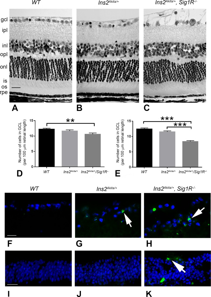
Figure 5.
Retinal structure of WT (Ins2+/+), Ins2Akita/+, and Ins2Akita/+/Sig1R−/−mice. Eyes of (A) WT (Ins2+/+), (B) Ins2Akita/+, and (C) Ins2Akita/+/Sig1R−/−mice were processed for JB-4 embedding and stained with H&E to permit visualization of retinal architecture. Animals were either 12 or 16 weeks at the time eyes were harvested. Panels shown are from 16-week-old mice. In WT (Ins2+/+) mice, the retinas are well organized and layers are of normal thickness. The retinas of the Ins2Akita/+ and Ins2Akita/+/Sig1R−/−mice are similar in appearance, with the exception of the nerve fiber layer in the Ins2Akita/+/Sig1R−/−mice (arrow), which is markedly disrupted. Retinas were subjected to morphometric analysis; there was a significant decrease in the number of cells in the GCL in the Ins2Akita/+/Sig1R−/−mice at 12 (D) and 16 weeks (E). Retinal cryosections of the same mice were subjected to immunofluorescent detection of TUNEL-positive cells (green labeling), cell nuclei were blue (DAPI) in (F) WT (Ins2+/+), (G) Ins2Akita/+, and (H) Ins2Akita/+/Sig1R−/−mice. Images were obtained using a high-powered objective focused on the GCL. Additional images of TUNEL staining were obtained using a lower-powered objective focused on the INL for (I) WT (Ins2+/+), (J) Ins2Akita/+, and (K) Ins2Akita/+/Sig1R−/−mice. gcl, ganglion cell layer; ipl, inner plexiform layer; inl, inner nuclear layer; opl, outer plexiform layer; onl, outer nuclear layer; is, inner segment; os, outer segment; rpe, retinal pigment epithelium. Calibration bar: 50 μm. Statistical significance: **P < 0.01, ***P < 0.001.

