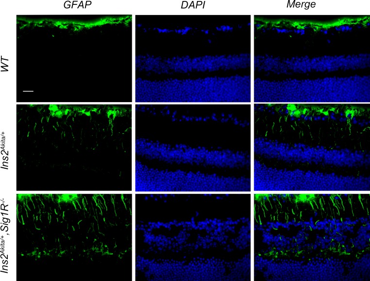
Figure 6.
Immunofluorescent detection of GFAP, a marker of gliosis in WT (Ins2+/+), Ins2Akita/+, and Ins2Akita/+/Sig1R−/−mice at 16 weeks. Representative photomicrographs of immunofluorescent detection of GFAP in retinal cryosections from (A) WT (Ins2+/+), (B) Ins2Akita/+, and (C) Ins2Akita/+/Sig1R−/−mice at age 16 weeks. Immunofluorescence analysis used anti-GFAP followed by incubation with Alexa Fluor 488 (green-labeled secondary antibody), showing marked increase in GFAP in radially oriented fibers of Müller glial cells (radial labeling) in Ins2Akita/+/Sig1R−/−mice but much less GFAP labeling in Müller cells of the WT or Ins2Akita/+ mice at this age. Astrocytic labeling with GFAP is typical and is observed in retinas of all mice. gcl, ganglion cell layer; inl, inner nuclear layer; onl, outer nuclear layer.

