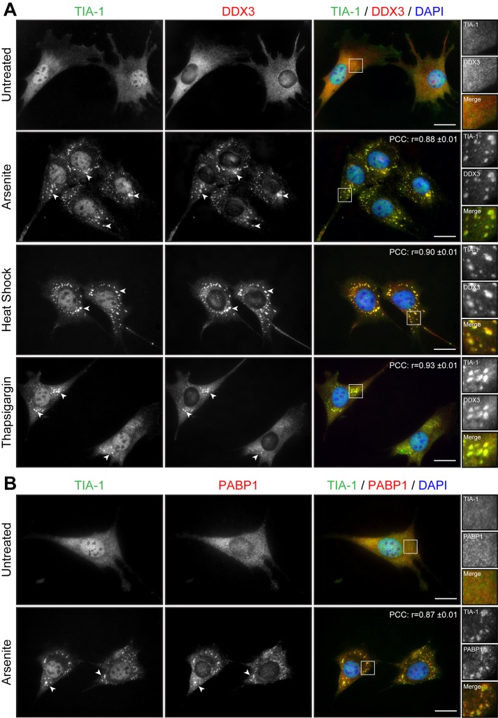
FIGURE 1:
Cell stress induces the formation of SGs in myoblasts. (A) Proliferative C2C12 myoblasts were untreated or treated with arsenite (0.5 mM for 45 min), by heat shock (45°C for 60 min), or with thapsigargin (1 μM for 60 min). Coimmunofluorescence stainings were performed using TIA-1 and DDX3 antibodies. (B) Coimmunofluorescence stainings were performed on untreated and arsenite-treated proliferative C2C12 using TIA-1 and PABP1 antibodies. DAPI was used to stain nuclei. Arrowheads show SGs in myoblasts. Right, magnifications of areas outlined by white boxes. Scale bars, 20 μm. For r, 40, 12, 11, and 11 cells were analyzed, respectively.

