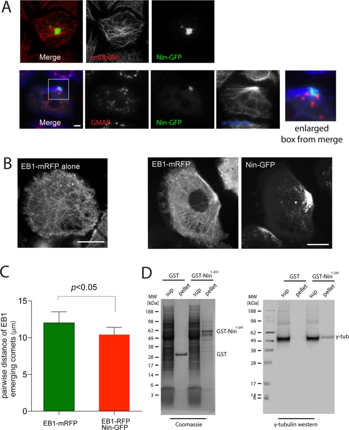
FIGURE 2:
Nin organizes microtubule-nucleating centers when overexpressed in Drosophila S2 cells. (A) Images of S2 cells expressing Nin-GFP. Microtubules are labeled with antibodies against α-tubulin, and Golgi with antibodies against GMAP. See also Supplemental Figure S1A. Scale bar, 5 μm. (B) Images of EB1-mRFP microtubule plus-end tracks in S2 cells with expression of Nin-GFP (bottom) or without (top). See also Supplemental Videos S1–S4. (C) Pairwise distance of EB1 emerging comets. Pattern of MT nucleation sites measured by plotting the point of emergence of each EB1 particle and correlating it with emergence of its neighbors. (D) GST-Nin N-terminal 241 amino acid domain binds to γ-tubulin in S2 cell lysates.

