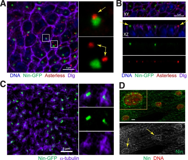
FIGURE 4:
Nin localizes to noncentrosomal MTOCs in wing epithelia and myocytes. (A) Nin-GFP associates with the noncentrosomal MTOC in wing epithelia. In the columnar epithelial cells of the developing wing disk, Nin-GFP (green) localizes primarily to one focus in each cell. The focus of Nin-GFP colocalizes adjacent to centrosomes ∼20% (29/156) of the time (top inset), and is unassociated in ∼80% (127/156) of cells (yellow arrows in insets). Centrosomes labeled with antibodies against asterless (asl), red). Dlg (purple) is an apical membrane marker. Image is an xy view of a third instar larval wing pouch epithelium z-stack projection. (B) Images of Nin-GFP foci in xy and xz views of the wing disk. These views demonstrate that Nin-GFP foci and centrosomes are both localized near the apical membrane in wing epithelia (yellow arrows). (C) Nin-GFP (green) localizes to the center of the noncentrosomal MTOCs labeled with α-tubulin (purple). (D) Myocytes in third instar larval muscles stained for endogenous Nin (green) using the C-terminal Nin antibody and DAPI (red) show that Nin has perinuclear localization. Yellow arrows point to the Nin localization at the periphery of myocyte nuclei. Scale bars: 5 μm in A–C, 1 μm in D.

