FIGURE 2:
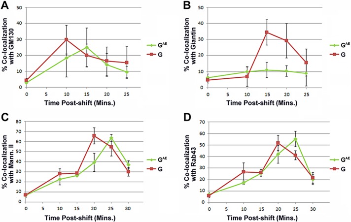
Quantification of GAE and G colocalization with various markers of the early secretory pathway in PH5CH8 cells. PH5CH8 hepatocytes expressing GAE-EGFP or G-EGFP were grown overnight at restrictive temperature and then shifted to permissive temperature for various times ranging from 0 to 30 min. At each time point, the cells were fixed and stained with antibodies specific for (A) GM130, (B) giantin, (C) mannosidase II, or (D) Rab43 and analyzed by confocal microscopy. The data points reflect percentage colocalization of GAE or G with the various early secretory pathway markers. The colocalization values shown represent the average value obtained from Z-stacks of at least 25 different cells from two independent experiments. Additional analyses revealed that the observed differences in the colocalization of G and GAE with giantin at the 15-min time point (p < 0.0001), with mannosidase II at the 20-min time point (p = 0.0002), and with endogenous Rab43 at the 25-min time point (p = 0.0006) were all statistically significant using the Student’s two-tailed t test.

