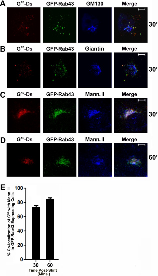
FIGURE 4:

GAE accumulates in a mannosidase II– and GFP-Rab43–containing compartment in GFP-Rab43–expressing cells. COS7 cells transfected with GFP-Rab43 were subsequently infected with adenovirus expressing GAE-Ds and grown at the restrictive temperature for 24 h. The cells were then shifted to permissive temperature for 30 min (A–C) or 60 min (D) and fixed. The cells were then permeabilized, stained with antibodies specific for GM130 (A), giantin (B), or mannosidase II (C, D), and incubated with Alexa Fluor 647–conjugated secondary antibody. The cells were then imaged by confocal microscopy. Scale bars, 5 μm. (E) Percentage colocalization of GAE-Ds with mannosidase II after a 30- or 60-min temperature shift. Average values from 20 different cells from two independent experiments.
