FIGURE 7:
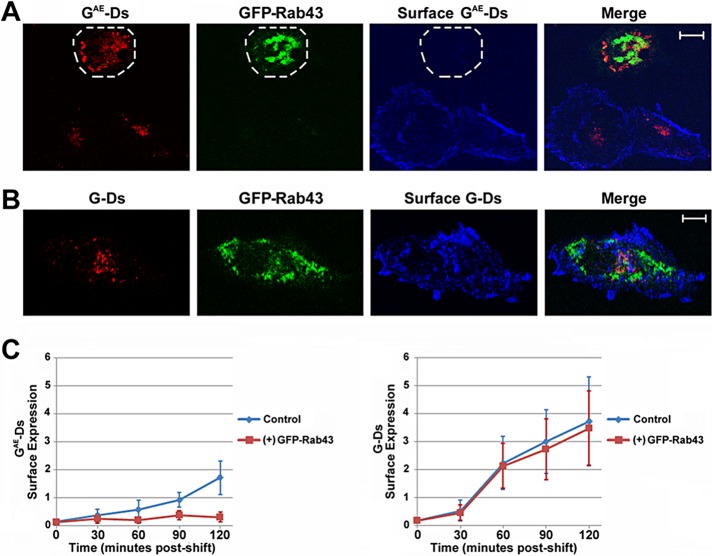
Differential effect of GFP-Rab43 on the surface accumulation of GAE and G. COS7 cells transfected with GFP-Rab43 were subsequently infected with adenoviral vectors expressing (A) GAE-Ds or (B) G-Ds and grown at the restrictive temperature for 24 h. The cells were then shifted to permissive temperature for 60 min and fixed. Surface GAE or G was stained in nonpermeabilized cells using the I1 monoclonal antibody, which recognizes an epitope in the ectodomains of both GAE and G, and an Alexa Fluor 647 secondary antibody. The cells were then imaged by confocal microscopy. The periphery of a cell coexpressing GFP-Rab43 and GAE-Ds is outlined in A. (C) Quantification of the surface levels of GAE-Ds and G-Ds in GFP-Rab43– expressing cells shifted to permissive temperature for times ranging from 0 to 120 min (averaged from 12–16 cells in two independent experiments). The difference in the surface delivery of GAE-Ds at the 120-min time point in the presence and absence of GFP-Rab43 is statistically significant, with p = 0.0003. Scale bars, 5 μm.

