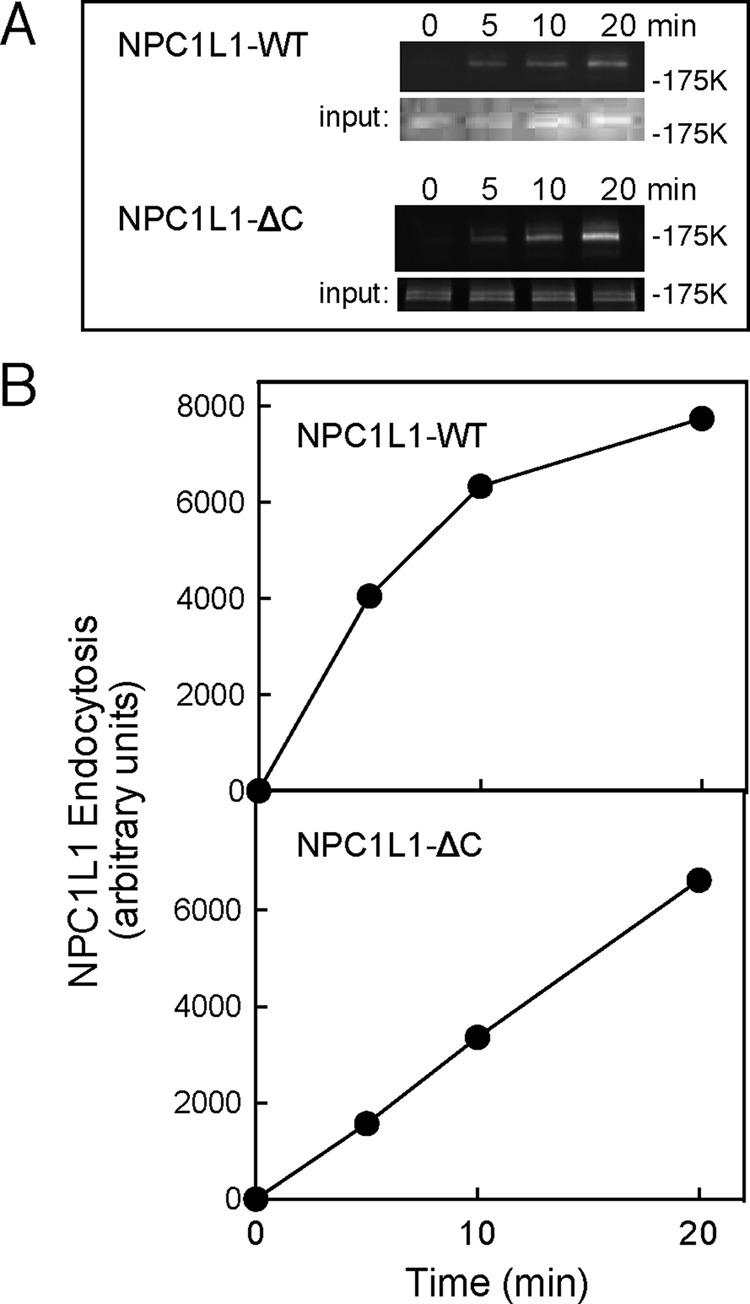
FIGURE 3:

Cell surface biotinylation assays of NPC1L1 endocytosis. RH7777 cells expressing NPC1L1-GFP or NPC1L1ΔC-GFP were surface labeled and assayed for endocytosis as in Figure 1. (A) Anti-GFP blots: input, total cell extract (8%); top, streptavidin-captured, surface-biotinylated proteins (40%). For NPC1L1-GFP, a representative experiment from six experiments carried out in duplicate is shown; the combined data were used for the normalized, control panels of Figures 4B and 5, B and C. Note that in this and the following experiments, most of the input represents internal NPC1L1; it provides a loading control for each sample, but the absolute signals cannot be compared with the endocytosis data, as they were obtained from separate gels. (B) Quantitation of data in A.
