Figure 2.
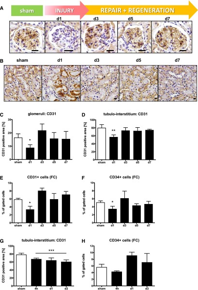
Endothelial injury and regeneration. Representative pictures of zinc fixed sections stained with anti-CD31 in glomeruli (A) and the tubulo-interstitium (B) (large scale=10 μm). Assessment of the glomerular (C) or peritubular endothelium (D) after staining for CD31 positive area measured using densitometry. Reduction of CD31 (E) and CD34 (F) positive cells measured by flow cytometry (fc) following selective EC injury or I/R (G, H).

